The Gut Wall Is a Controlled Border
Digestion is only preparation. Nutrients do not become available to the rest of the body until they cross the intestinal lining—a living border that decides what can enter, how it crosses, and what must remain in the digestive tract.
This crossing is called absorption. It happens mainly in the small intestine, whose structure is built to combine enormous surface area with tight control.
The key idea is selectivity. The gut wall is not an open net that lets every small object through. Its cells recognize, transport, package, and sometimes modify nutrients as they enter.
Where the Meal Is Now
Return to the lentils, rice, vegetables, yogurt, and oil from the opening article. By the time this mixture reaches the small intestine, it no longer looks much like the original plate. Starch and protein are being dismantled, fat is dispersed into smaller droplets, and water surrounds the dissolved material. Yet almost all of it is still on the digestive-tract side of the border. The next decisive event is crossing.
The Small Intestine Creates a Huge Contact Surface
Efficient absorption requires contact between digested food and intestinal cells. The small intestine creates that contact by folding its inner surface again and again.
Large circular folds line the intestine. On those folds sit millions of tiny finger-shaped projections called villi. Each villus is covered by intestinal cells whose own surface has an even finer brush-like border called microvilli.
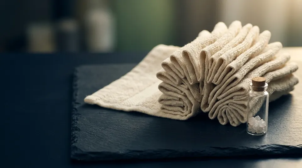
A useful analogy is a flat towel compared with a deeply folded one. Both fit in the same space, but the folded towel exposes far more material. Villi and microvilli give nutrients far more surface on which to meet an absorptive cell.
Inside each villus are small blood vessels and a lymph vessel. Once a nutrient crosses the intestinal cells, one of these transport routes is waiting nearby.
Digestion Produces Pieces the Border Can Handle
Large food molecules generally cannot cross the intestinal lining intact. Digestion dismantles them into smaller units first.
Carbohydrate digestion produces single sugars, including glucose, fructose, and galactose. Protein digestion produces amino acids and very small chains of amino acids. Fat digestion produces fatty acids and smaller fat components.
The sequence matters. Enzymes break a large molecule into absorbable pieces. Those pieces reach the surface of an intestinal cell. A suitable route then moves them into or through that cell.
Fiber behaves differently because human digestive enzymes cannot dismantle many of its chemical bonds. Most fiber therefore remains in the digestive tract and continues to the large intestine, where gut microbes can use some types.
Different Nutrients Use Different Doors
The intestinal lining uses several ways to move substances across. The simplest is diffusion: a substance moves from an area where it is more concentrated toward an area where it is less concentrated.
Some nutrients cannot pass through the cell’s outer membrane on their own. They use a protein embedded in the membrane as a doorway. When the movement still follows the concentration difference, this is called facilitated diffusion—facilitated because a transport protein helps.
Other nutrients must be moved against a concentration difference, from where there is less toward where there is more. That requires energy, directly or indirectly, and is called active transport.
Glucose shows how these methods can work in sequence. At the gut-facing side of an intestinal cell, a transport protein brings glucose in together with sodium. The cell maintains the sodium difference using energy, so that sodium movement can pull glucose along.
Glucose then leaves the other side of the intestinal cell through another transport protein and enters nearby fluid and blood. One doorway brings it into the border cell; another lets it out toward the body.
Fructose uses a different transport protein and does not rely on the same sodium-linked entry step. The two sugars may look similar on a food label, but their first steps across the intestine are not identical.
Water Follows Dissolved Substances
Water absorption is closely tied to the movement of salts and nutrients. Water tends to move toward the side with more dissolved particles, a process called osmosis.
As sodium, glucose, amino acids, and other substances move through the intestinal lining, water follows. This is why the intestine can absorb large amounts of fluid from drinks, food, and its own digestive secretions.
The same principle explains oral rehydration solutions used during severe fluid loss. A carefully balanced mixture of glucose and sodium uses their shared intestinal transport route; as both are absorbed, water follows. More sugar is not better—the concentrations must be appropriate for the system to work well.
Fat Has to Be Repackaged
Most dietary fat cannot simply dissolve in the watery contents of the intestine or in blood. It needs temporary carriers and new packaging.
Bile breaks large fat droplets into smaller droplets, giving digestive enzymes more surface to work on. Enzymes then release fatty acids and other small fat components.
Bile molecules gather these components into tiny transport clusters that can approach the intestinal cell. The fat components leave the cluster and enter the cell.
Inside, the cell rebuilds much of the fat and packages it with protein into a transport particle called a chylomicron. Chylomicrons are too large for the small blood vessels in a villus, so they enter the nearby lymph vessel instead.
Fat-soluble vitamins—A, D, E, and K—generally travel with this fat-handling system. This is why some dietary fat helps their absorption. It does not mean more fat always produces more absorption; enough to support the process is what matters.
The Barrier Has to Admit Nutrients and Exclude Threats
The intestinal lining performs two jobs that pull in opposite directions. It must allow a large daily flow of nutrients and water through while limiting entry by microbes, toxins, and unwanted large molecules.
Neighboring intestinal cells are joined by protein structures called tight junctions. These junctions regulate the space between cells. A mucus layer, immune cells, digestive secretions, and the rapid replacement of lining cells add further protection.
The barrier is dynamic, not a brick wall. Infections, inflammation, alcohol, some medicines, and certain diseases can disturb it. But popular “leaky gut” claims often jump from a real biological concept to broad explanations for vague symptoms without adequate evidence.
For everyday health, the useful point is simpler: a healthy intestine is selectively permeable. It is designed to let the right materials cross, not to be completely sealed.
Absorption Is Efficient, but Not Identical for Every Nutrient
A food label tells you what the product contains, not exactly what every person will absorb. The proportion available for absorption is often called bioavailability.
Chemical form matters. Iron from animal foods and iron from plants use partly different pathways and are absorbed at different rates. Meal composition matters too: vitamin C can improve absorption of plant iron, while some plant compounds can reduce it.
Body need can also matter. The intestine adjusts absorption of some nutrients according to stores and hormonal signals. Vitamin D, for example, helps regulate calcium absorption.
These differences are real, but they do not mean every meal needs to be engineered for maximum uptake. A varied dietary pattern gives the body repeated opportunities to absorb what it needs, and excessive absorption would not always be beneficial.
The Main Idea
The intestinal wall is the point where digested food becomes available to the internal body. Folds, villi, and microvilli create surface area; digestive enzymes create absorbable pieces; and specialized transport routes move different nutrients across.
Keep one distinction clear: digestion breaks food down; absorption moves it across. The next step is routing—why many nutrients travel directly to the liver while most dietary fat begins its journey through lymph.